
The DICOM Viewer
A powerful, friendly multi-modality DICOM viewer
MODALIZER+’s DICOM Viewer displays images, video, structured reports and encapsulated PDFs from any modality, side by side, with a full set of measurement, contrast, and editing tools. This guide covers every viewer feature in detail. For the free, always-available version, see the Free DICOM Viewer page.
Viewer Layout
- Main display area – shows the selected image. Press Ctrl+H to show/hide DICOM attributes, Ctrl+T to show/hide the text overlay. Right-click for a context menu with tool controls, sending the image to PACS, and saving it in its original SOP class/transfer syntax or as JPEG/PNG/BMP/PDF/MPEG – every file can also be saved as a text DICOM dump. Press F11 for full screen (Esc or the on-screen ‘x’ to exit).
- Patients and Studies tree – right-click a study row to send it to PACS, close it, or export it to external media (Local Archive studies only).
- Series thumbnails for the selected study – right-click a thumbnail to send that series to PACS.
- Image previews within the selected series, shown as thumbnails along the bottom. Spatially related images are grouped rather than shown individually. Click a thumbnail, or use the left/right arrows, to load the next or previous image; right-click an image to send just that one to PACS.
- Tool buttons strip for viewer operations and loading new images from a folder, file dialog, or history – every tool is available from the toolbar or the right-click context menu.
Every panel can be expanded or collapsed using the arrow icon at its top-left corner. Right-click the toolbar itself to customize which tool groups are visible.
Loading Images into the Viewer
Drag-and-drop files onto the viewer, click Open Files, or use Ctrl+O to browse for a folder. Use Open Folder to load an entire folder’s content at once, or open the History screen with the History button or Ctrl+Shift+H.
Each open action (Files, Folder, DICOMDIR, etc.) lists one or more study rows, formatted as “[Patient Name], [Patient ID], [Accession Number]”. Type in the Search field to find a study quickly, then click Open selected item to load it.
DICOM files with embedded video play in the built-in video player (or your system player); embedded PDFs open in your system PDF viewer; SR reports open in the built-in HTML viewer.
Split-Screen: Up to Four Studies at Once
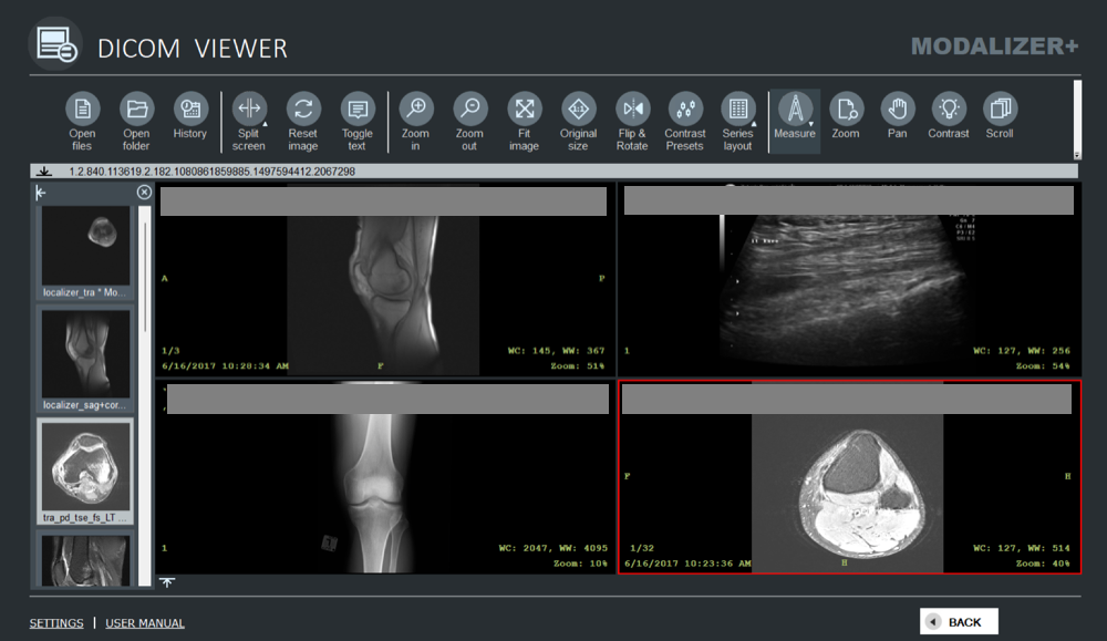
Use the split options in the toolbar to display up to four studies side by side simultaneously – useful for comparing prior and current studies, or different modalities, in one view. Click a section to highlight it (a red frame appears), then click a study or series thumbnail – or simply drag a thumbnail into the section where you want it displayed.
Hounsfield Unit Contrast Presets
MODALIZER+ ships with a set of predefined contrast presets, available from the “Contrast presets” tool button, to instantly change the window center (WC) and window width (WW) values of the current image – a fast way to switch between bone, lung, soft-tissue and other Hounsfield unit windows on CT studies.
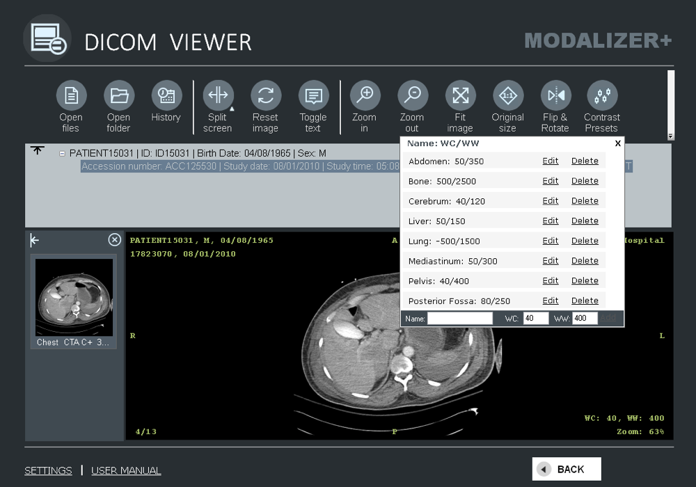
Add a custom preset by filling in Name/WC/WW values and clicking Add; existing presets can be edited or deleted from the same pop-up. If an image contains one or more VOI LUTs (Values of Interest Lookup Tables), those are automatically added to the preset list, and the first VOI LUT pair is applied when the image opens.
Measurement and Annotation Tools
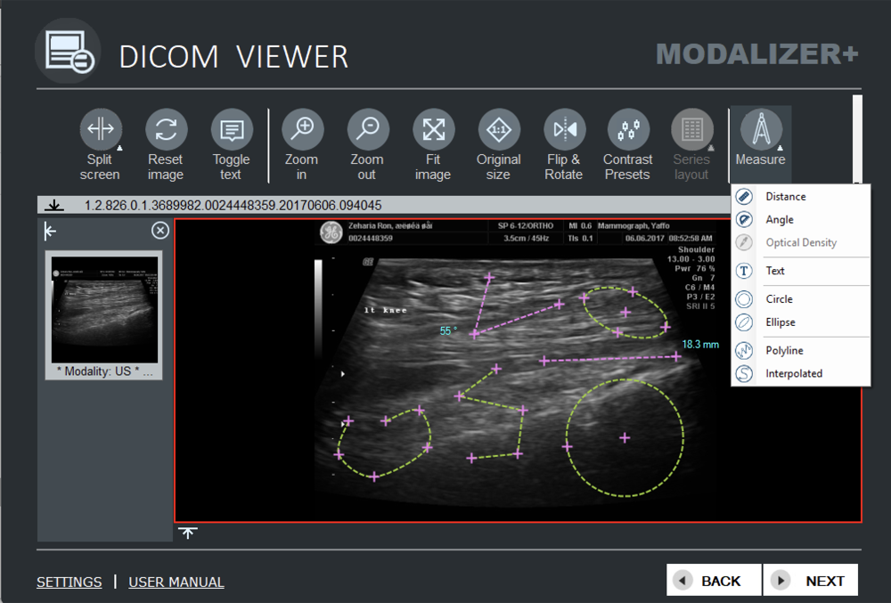
The DICOM Viewer supports every graphic annotation type defined in the DICOM standard:
- Distance (length in mm)
- Angle (degrees)
- Optical Density (Hounsfield Units)
- Text annotation
- Circle (diameter in mm and area in mm²)
- Ellipse/Oval (major/minor axis diameters in mm and area in mm²)
- Polyline
- Interpolated line
All annotations are stored per the DICOM standard and can be displayed by any supporting DICOM viewer. Click once to start a distance or angle measurement, click again to place each subsequent point – the live measurement updates as you move the mouse. Click once for optical density. To edit a measurement, grab an endpoint or the whole line and drag it; select any part of a line and press Delete to remove it.
You can save measurements as a Grayscale Softcopy Presentation State (GSPS) file under a new series. When closing the viewer with unsaved annotations, you’ll be prompted to:
- Add the new items to the Local Archive (if enabled) – if the study is already archived, only the new GSPS file is added.
- Send the entire study, or just the new files, to PACS.
- Save the new GSPS file to a folder of your choice.
- Discard the changes without saving.
12-Lead ECG Viewer
MODALIZER+ can display DICOM 12-Lead ECG instances directly in the viewer.
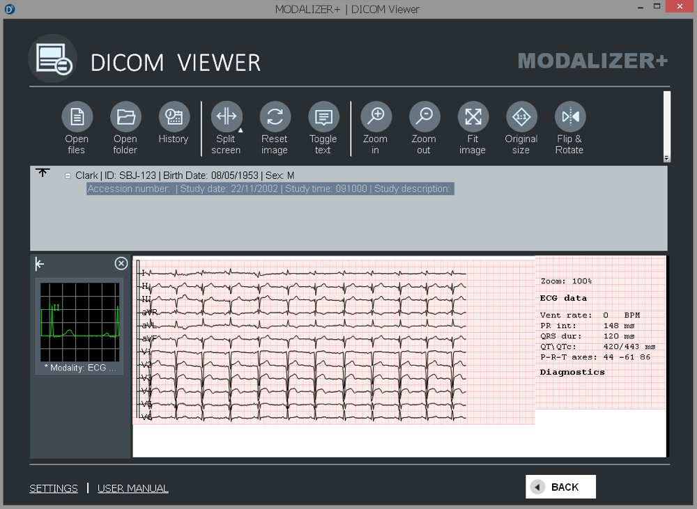
Use the ECG Display Settings tab in Settings to choose the graph format: click “Settings” from the Viewer, adjust the parameters, and click “Back” – the new display settings apply immediately.
Zoom, Pan, Contrast and Scroll Tools
Click any of these tool buttons to activate it – the button background turns red and the mouse cursor changes to match. Press and hold the left mouse button while moving the pointer to zoom, pan the image, adjust window/level contrast, or scroll through frames, depending on the active tool.
Flip and Rotate
Available actions: Flip Vertically, Flip Horizontally, Rotate 90° Clockwise, and Rotate 90° Counterclockwise. Any measurements on the current image are transformed to match.
CINE Tool for Multi-Frame Playback
When a loaded DICOM file contains a multi-frame image, CINE mode activates automatically and the CINE controls appear.
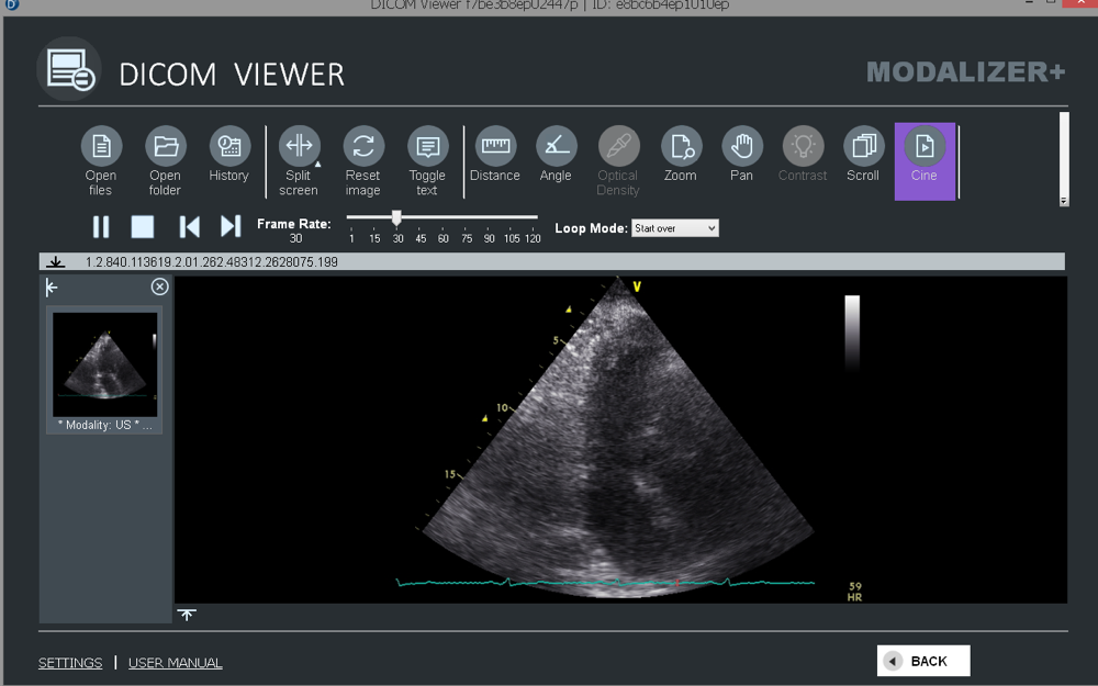
- Click the Play/Pause button to start or pause CINE playback.
- Click the Stop button to stop CINE and close the CINE panel.
- Use the first/last frame buttons to jump to either end of the sequence.
- Adjust the CINE frame rate and loop mode directly from the CINE panel.
Grayscale Softcopy Presentation State (GSPS)
MODALIZER+ displays graphic and text annotations stored in DICOM Presentation State objects (GSPS). When you open a study, the viewer automatically loads every GSPS file’s data – the relationships between annotations and study instances/frames, annotation types and positions – and converts it into graphic objects drawn over the matching images.
Each GSPS module (a series with PR modality) appears as its own thumbnail in the left panel, just like any other series. Selecting a PR thumbnail displays its graphic objects as separate image thumbnails at the bottom, with the first one shown in the main area over the image/frame it references. Selecting a non-PR series hides the annotations. With a PR series selected, choosing “Show header” displays its tags (editing is not available for PR files).
DICOM Header View
Right-click any image and select “Show header”, or press Ctrl+H, to view every data element in the DICOM file. Use the search bar at the top of the header view to find elements by tag number, tag name, or value.
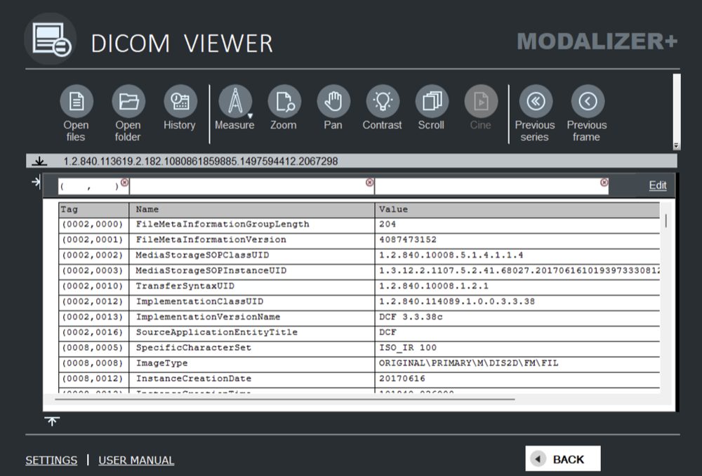
By default, private tags display with a “?” name – you can configure private tag names and types using the dicom.dic file in the installation folder (tab-separated columns: Tag, VR, Name, VM, Version). The installed dicom.dic includes a few example lines; edit or delete them in Notepad as needed.
DICOM Editor – a Built-in DICOM Q/A Workstation
MODALIZER+ doubles as a full DICOM Q/A workstation, with editing available at the Instance, Series, or Study level, IHE IOCM-compliant, with every change reviewable before it’s committed. This is a major enough feature to warrant its own chapter – see the full DICOM Editor guide for entering edit mode, what you can edit, and how the review and IHE IOCM workflow works.
Image Info View
Select “Image Info” from the context menu to see the displayed image’s metadata – size, compression method, and color space.
DICOM Viewer Shortcut Keys
| Shortcut | Action |
|---|---|
| Ctrl + H | Show/Hide text overlay and DICOM header |
| Ctrl + O | Open File |
| Ctrl + Shift + H | Show History |
| F11 | Full screen mode |
| Esc | Exit full screen mode |
← Previous: Quick Start Guide | Next: DICOM Editor →
DICOM is the registered trademark of the National Electrical Manufacturers Association for its standards publications relating to digital communications of medical information.